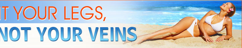
varicose-help.comvaricose-help.com

DVT & Pulmonary Embolism

Varicose veins can be much more than a superficial situation, marked by the bulging and twisted veins that appear on the legs and sometimes in the groin area. Varicose veins can be a precursor to deep vein thrombosis, or DVT. A blood clot, which occurs when blood thickens and clumps together, forms in a vein deep inside the body. Such clots usually form in the leg or thigh, but are not restricted to those areas.

When DVT Becomes PE

In deep vein thrombosis, a blood clot in a deep vein (called an embolus) can break off and travel through the bloodstream. The popular belief has held that the blood clot ends up in the heart, but this is not the case. The clot travels through the right side of the heart and lodges in the lungs, reducing the amount of oxygen that enters the body. When one such clot arrives to the lungs and blocks the flow of blood, the condition is called pulmonary embolism (PE). It is one of the most feared complications of DVT and is the third most common cause of death in the US. It is also the highest cause of preventable deaths in hospital patients.

Left Undiagnosed, It Can Kill

The unnerving thing about pulmonary embolism is that it can occur without any signs or symptoms. Sometimes the body is able to handle the clot adequately and no symptoms appear. The well-known and classical symptoms of PE include a sudden sharp, stabbing pain in the chest, tenderness of the chest area, back and/or shoulder pain. Breathing pain and coughing up blood can also be symptoms of PE. There may also be a change in the rhythm of the heartbeat. When a physical examination is done, the doctor will often find low blood pressure, fast breathing or tenderness in the chest wall. Since these symptoms are often quite general, it often takes far more indications for a diagnosis to be made. In the meantime, people with undiagnosed PE who do not receive any type of treatment have a high incidence of recurring pulmonary embolism and death.

Making The Diagnosis And Treating PE

Diagnosis is achieved through testing. The favored method used today is the CT chest scan using an IV contrast. The PE will show up as a dark spot in the veins of the lungs. If a more intensive evaluation is necessary, then an invasive test can be done. Once the presence of PE is established, the aim is then to stabilize the person and plan intervention. In most cases, blood thinners are used to dilute the consistency of the blood in order to promote healing in the lungs.

Some people cannot handle blood thinners and an alternative treatment must be established. In such cases, an inferior vena cava (IVC) filter is placed, using x-ray guidance, under the kidney veins in the vena cava. The IVC drains both legs in order to prevent any other blood clots from travelling up to the lungs. If treatment that is more aggressive is needed, the option of removing or dissolving the clot is considered using mechanical aids and dissolving medications. Since there is a very high mortality rate with surgical removal, this option is least used.